| Endocrine Index | Glossary |
|---|
 |
Insulin Synthesis and Secretion |
|
| |
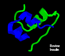
Structure of InsulinInsulin is a rather small protein, with a molecular weight of about 6000 Daltons. It is composed of two chains held together by disulfide bonds. The figure to the right shows a molecular model of bovine insulin, with the A chain colored blue and the larger B chain green. You can get a better appreciation for the structure of insulin by manipulating such a model yourself. The amino acid sequence is highly conserved among vertebrates, and insulin from one mammal almost certainly is biologically active in another. Even today, many diabetic patients are treated with insulin extracted from pig pancreas. |
 |
Biosynthesis of InsulinInsulin is synthesized in significant quantities only in B cells in the pancreas. The insulin mRNA is translated as a single chain precursor called preproinsulin, and removal of its signal peptide during insertion into the endoplasmic reticulum generates proinsulin.
When the B cell is appropriately stimulated, insulin is secreted from the cell by exocytosis and diffuses into islet capillary blood. C peptide is also secreted into blood, but has no known biological activity. Control of Insulin SecretionInsulin is secreted in primarily in response to elevated blood concentrations of glucose. This makes sense because insulin is "in charge" of facilitating glucose entry into cells. Some neural stimuli (e.g. sight and taste of food) and increased blood concentrations of other fuel molecules, including amino acids and fatty acids, also promote insulin secretion. Our understanding of the mechanisms behind insulin secretion remain somewhat fragmentary. Nonetheless, certain features of this process have been clearly and repeatedly demonstrated, yielding the following model:
The figure to the right depicts the effects on insulin secretion when enough glucose is infused to maintain blood levels two to three times the fasting level for an hour. Almost immediately after the infusion begins, plasma insulin levels increase dramatically. This initial increase is due to secretion of preformed insulin, which is soon significantly depleted. The secondary rise in insulin reflects the considerable amount of newly synthesized insulin that is released immediately. Clearly, elevated glucose not only simulates insulin secretion, but also transcription of the insulin gene and translation of its mRNA.
|
| Index of: The Endocrine Pancreas | |||
|---|---|---|---|
 |
Functional Anatomy of the Endocrine Pancreas | Physiologic Effects of Insulin |  |
|
Last updated on June 15, 1999 |
| Author: R. Bowen |
| Send comments via form or email to rbowen@colostate.edu |